NLS Analysis of Bone Marrow and Postoperative Spinal Changes
NLS imaging technology provides a digital approach to observing structural patterns and changes in spinal tissues. When examining areas surrounding degenerative intervertebral discs, bone marrow patterns may be described in three general categories: vascular, fatty, and sclerotic.
A comprehensive NLS examination may include several scanning approaches to observe different anatomical areas and tissue patterns.
1. Two-Dimensional and Three-Dimensional Scanning
Two-dimensional scanning in sagittal and axial projections helps visualize the areas surrounding affected intervertebral discs. These views provide different perspectives for examining disc-related structural patterns.
Three-dimensional scanning can provide additional visualization of the vertebral endplates and adjacent bone marrow regions. It may help highlight areas of interest, including changes in endplate appearance and surrounding tissue patterns.
2. NLS Microscanning for Spinal Structures
NLS microscanning offers a more detailed approach to examining selected spinal regions. It can be used to observe structural patterns around the dural sac and nerve root channels, including areas of narrowing, deformation, or positional changes.
These observations may provide additional reference information when reviewing spinal structural changes.
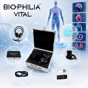
3. Monitoring Postoperative Changes with Biophilia Vital
With its non-invasive operating approach and lack of ionizing radiation, Biophilia Vital can be used for repeated observational assessments of selected tissue patterns following spinal surgery.
Spectral-Entropy Analysis (SEA) is another analytical feature used to compare different signal patterns. In postoperative observations, it may help distinguish between patterns associated with disc tissue and scar tissue.
However, NLS and spectral-entropy analysis should be considered complementary observational tools rather than clinically validated methods for diagnosing recurrent disc herniation or determining postoperative complications.
Conclusion
Biophilia Vital combines NLS-based scanning approaches and spectral-entropy analysis to support the observation of spinal tissue patterns. Its non-invasive approach makes it suitable for repeated informational assessments, while conventional medical imaging and professional clinical evaluation remain essential for diagnosing spinal conditions and assessing postoperative outcomes.